Anemia
From Wikipedia, the free encyclopedia
For other uses, see Anemia (disambiguation).
| Anemia | |
|---|---|
| Classification and external resources | |
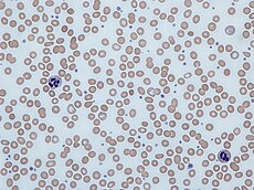
 Human blood from a case of iron-deficiency anemia |
|
| ICD-10 | D50-D64 |
| ICD-9 | 280-285 |
| DiseasesDB | 663 |
| MedlinePlus | 000560 |
| eMedicine | med/132 emerg/808 emerg/734 |
| MeSH | D000740 |
Anemia is the most common disorder of the blood. The several kinds of anemia are produced by a variety of underlying causes. It can be classified in a variety of ways, based on the morphology of RBCs, underlying etiologic mechanisms, and discernible clinical spectra, to mention a few. The three main classes include excessive blood loss (acutely such as a hemorrhage or chronically through low-volume loss), excessive blood cell destruction (hemolysis) or deficient red blood cell production (ineffective hematopoiesis).
Of the two major approaches to diagnosis, the "kinetic" approach involves evaluating production, destruction and loss,[3] and the "morphologic" approach groups anemia by red blood cell size. The morphologic approach uses a quickly available and low-cost lab test as its starting point (the MCV). On the other hand, focusing early on the question of production may allow the clinician to expose cases more rapidly where multiple causes of anemia coexist.
Contents |
Signs and symptoms

Main symptoms that may appear in anemia[4]
Most commonly, people with anemia report feelings of weakness, or fatigue, general malaise and sometimes poor concentration. They may also report dyspnea (shortness of breath) on exertion. In very severe anemia, the body may compensate for the lack of oxygen-carrying capability of the blood by increasing cardiac output. The patient may have symptoms related to this, such as palpitations, angina (if pre-existing heart disease is present), intermittent claudication of the legs, and symptoms of heart failure.
On examination, the signs exhibited may include pallor (pale skin, mucosal linings and nail beds), but this is not a reliable sign. There may be signs of specific causes of anemia, e.g., koilonychia (in iron deficiency), jaundice (when anemia results from abnormal break down of red blood cells — in hemolytic anemia), bone deformities (found in thalassemia major) or leg ulcers (seen in sickle-cell disease).
In severe anemia, there may be signs of a hyperdynamic circulation: tachycardia (a fast heart rate), bounding pulse, flow murmurs, and cardiac ventricular hypertrophy (enlargement). There may be signs of heart failure.
Pica, the consumption of non-food items such as soil, paper, wax, grass, ice, and hair, may be a symptom of iron deficiency, although it occurs often in those who have normal levels of hemoglobin.
Chronic anemia may result in behavioral disturbances in children as a direct result of impaired neurological development in infants, and reduced scholastic performance in children of school age. Restless legs syndrome is more common in those with iron-deficiency anemia.
Diagnosis

Peripheral blood smear microscopy of a patient with iron-deficiency anemia
In modern counters, four parameters (RBC count, hemoglobin concentration, MCV and RDW) are measured, allowing others (hematocrit, MCH and MCHC) to be calculated, and compared to values adjusted for age and sex. Some counters estimate hematocrit from direct measurements.
| Age or gender group | Hb threshold (g/dl) | Hb threshold (mmol/l) |
|---|---|---|
| Children (0.5–5.0 yrs) | 11.0 | 6.8 |
| Children (5–12 yrs) | 11.5 | 7.1 |
| Teens (12–15 yrs) | 12.0 | 7.4 |
| Women, non-pregnant (>15yrs) | 12.0 | 7.4 |
| Women, pregnant | 11.0 | 6.8 |
| Men (>15yrs) | 13.0 | 8.1 |
If an automated count is not available, a reticulocyte count can be done manually following special staining of the blood film. In manual examination, activity of the bone marrow can also be gauged qualitatively by subtle changes in the numbers and the morphology of young RBCs by examination under a microscope. Newly formed RBCs are usually slightly larger than older RBCs and show polychromasia. Even where the source of blood loss is obvious, evaluation of erythropoiesis can help assess whether the bone marrow will be able to compensate for the loss, and at what rate.
When the cause is not obvious, clinicians use other tests: ESR, ferritin, serum iron, transferrin, RBC folate level, serum vitamin B12, hemoglobin electrophoresis, renal function tests (e.g. serum creatinine).
When the diagnosis remains difficult, a bone marrow examination allows direct examination of the precursors to red cells.


0 komentar:
Posting Komentar